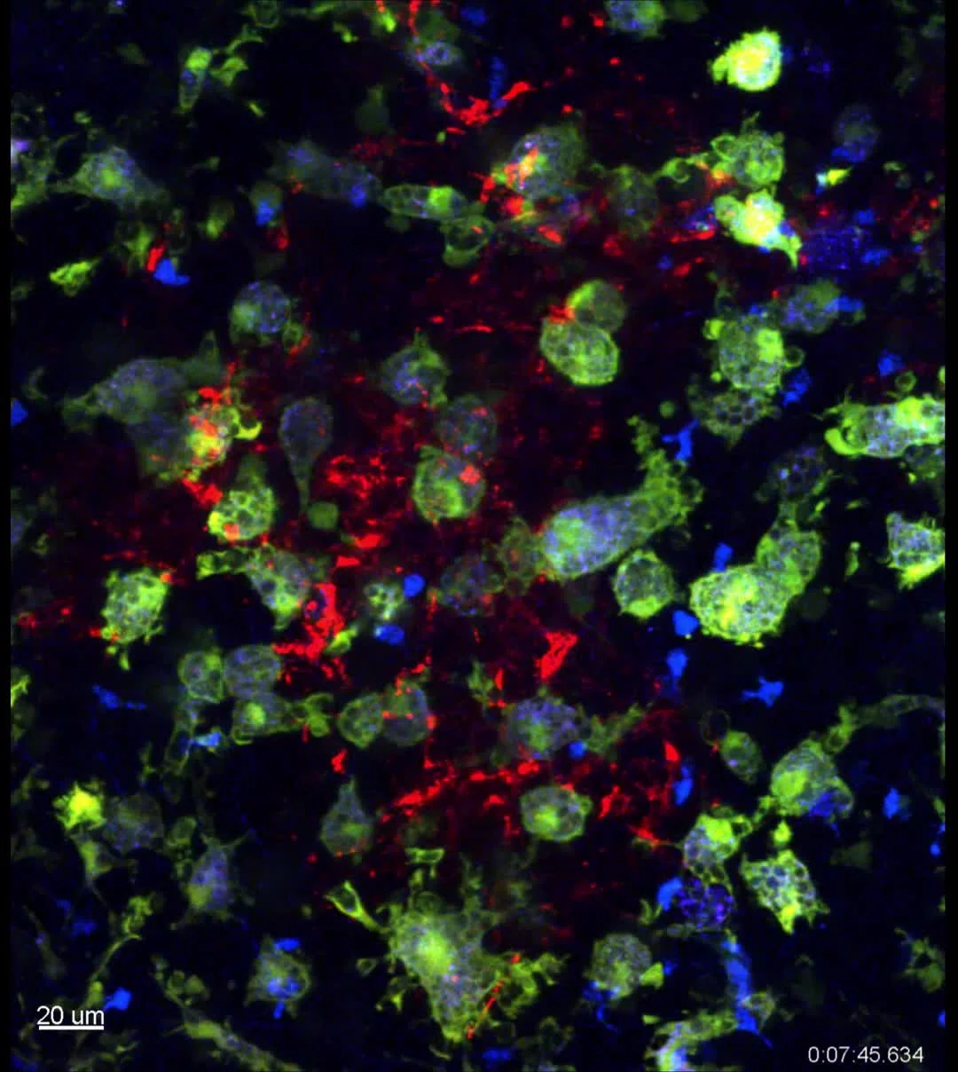
Foxp3+ T cells (blue) engage with CD11c+ Macropahges (green) in the germinal center.
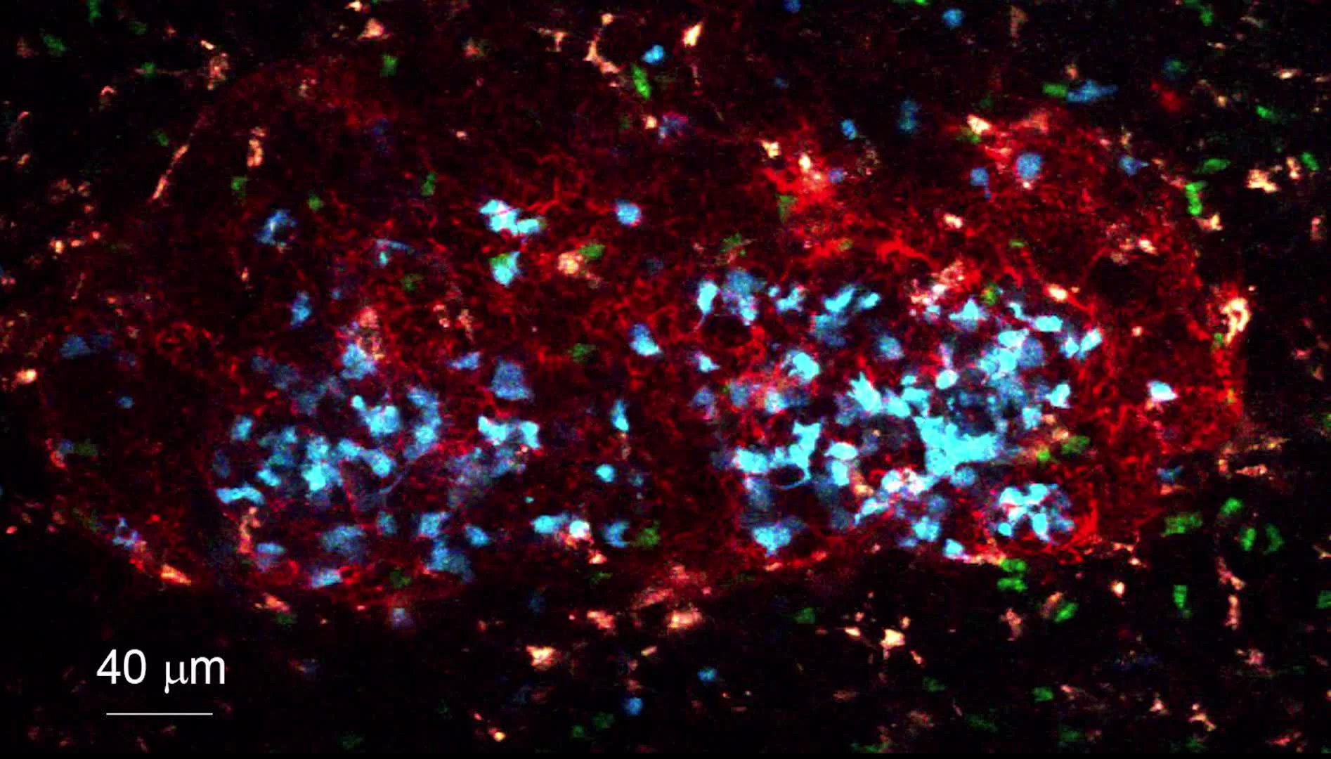
With a widow implanted on the inguinal lymph node we can image the collapse of a germinal center. Antigen specific B cells (blue), Foxp3+ T cells (green) and follicular dendritic cells (red) can be visualized as the germinal center collapses.
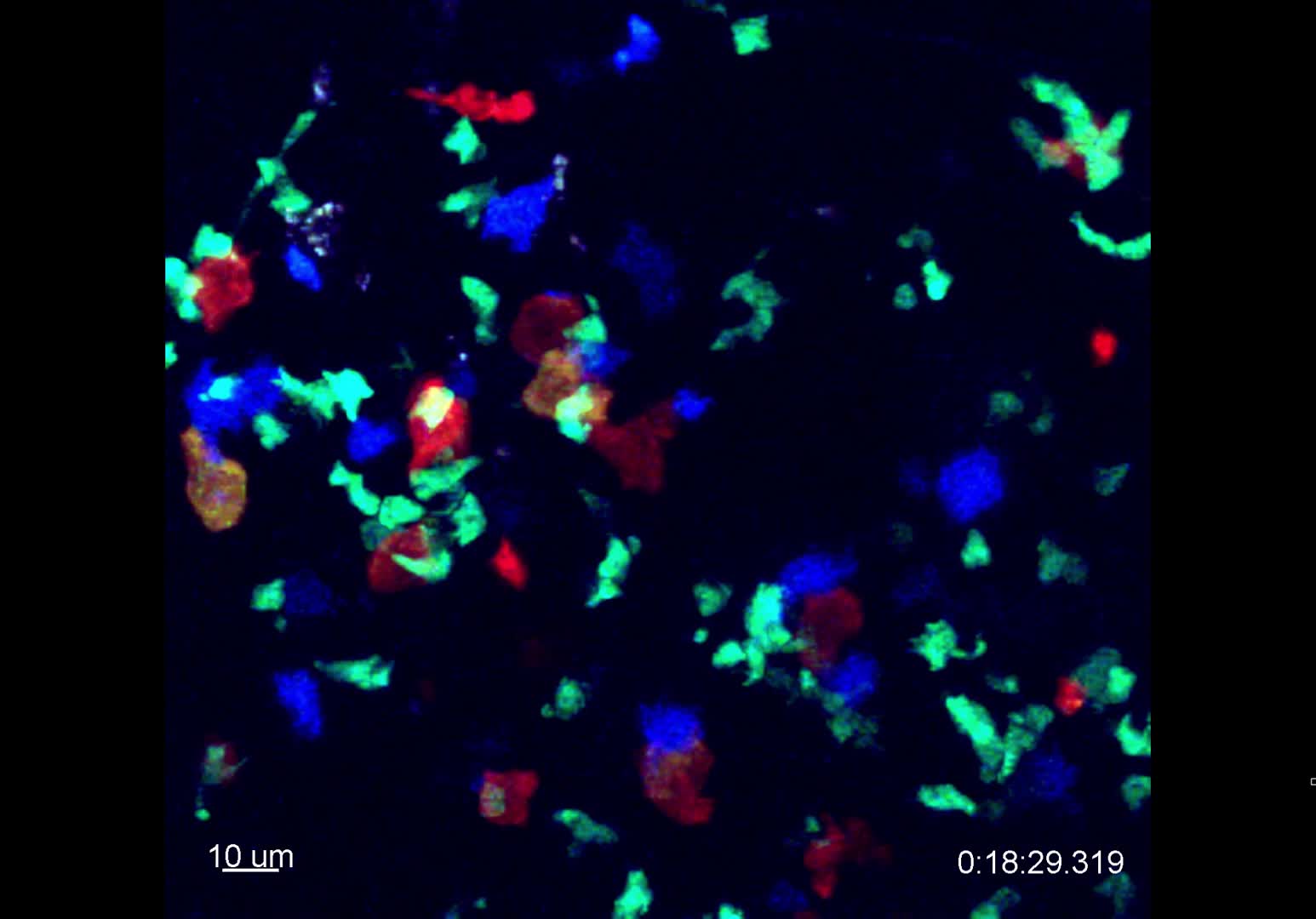
Antigen specific B cells (blue) interacts vigorously with antigen specific T cells (red) and Foxp3+ T cells (green) prior to germinal center formation. Signaling in antigen specific T cells can be visualized as flashing (orange) by a Calcium reporter.
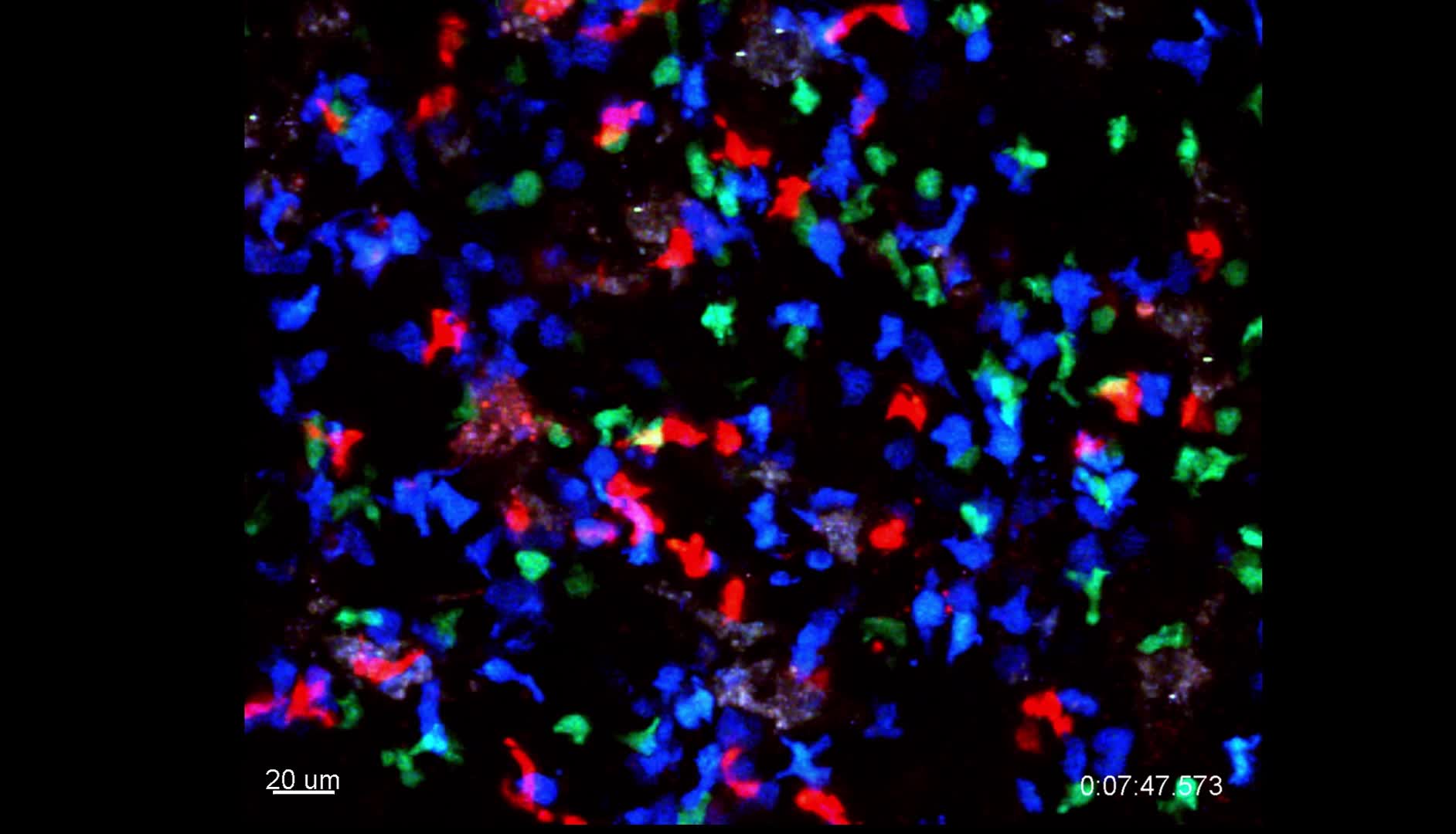
In collapsing germinal centers, we can observe interactions between antigen specific T cells (red) and germinal center B cells (blue), but also between Foxp3+ T cells (green) and germinal center B cells.

Antigen specific T cells (red) interact with germinal center B cells (blue), at the peak of the germinal center reaction. However, at this time point, Foxp3+ T cell (green) interactions with germinal center B cells can barely be detected.