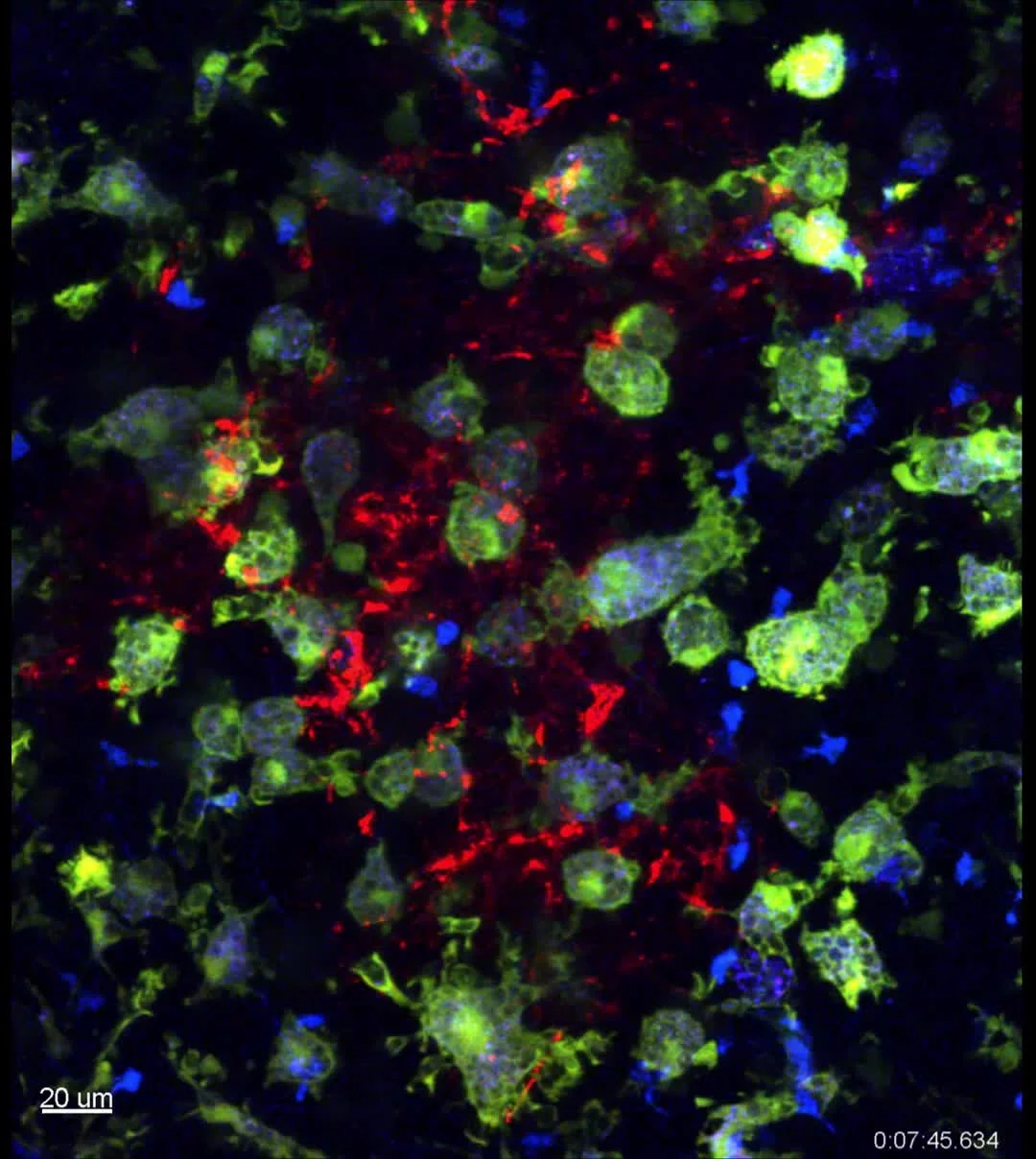
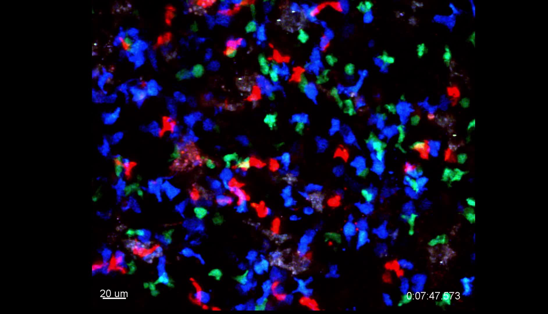
Longitudinal window imaging of a collapsing GC
With a widow implanted on the inguinal lymph node we can image the collapse of a germinal center. Antigen specific B cells (blue), Foxp3+ T cells (green) and follicular dendritic cells (red) can be visualized as the germinal center collapses.
Previous
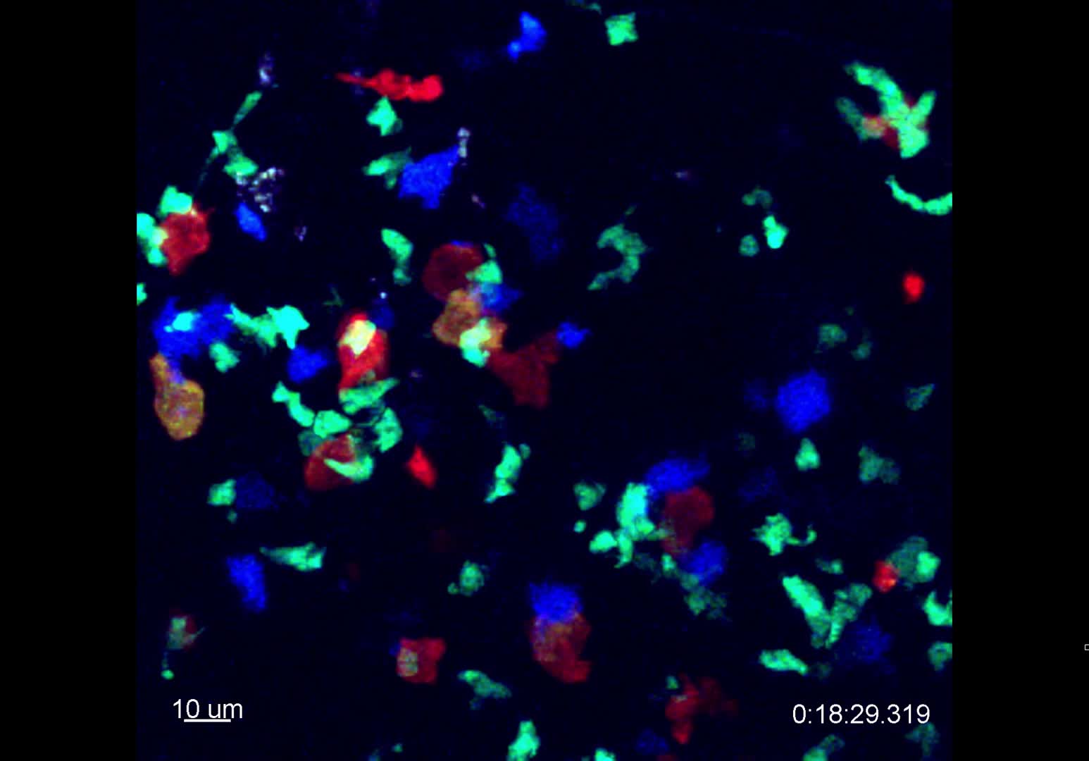
Foxp3+ T cells interacting with Macrophages in the GC
Next